Silicon Neural Probe Optogenetics

Photoelectric artifact-free electrophysiology combined with optogenetics in acute and freely-behaving animals

Enjoy artifact-free data with our silicon neural probes during optogenetic stimulation - a major breakthrough for combined electrophysiology with optogenetics.


Photoelectric artifact-free electrophysiology - why it matters...

A common problem when combining electrophysiology and optogenetics is that ideally you want your specific target neurons to respond to light but so too will your electrodes since when light strikes a metal electrode it commonly causes a photoelectric artifact due to the Becquerel effect. These artifacts may be large amplitude and long-lasting, resulting in potential data loss and / or contamination of your signal with artifacts that may mimic spikes so single unit recordings during optogenetic stimulation have proved tricky so far....examples of this in the literature can be found in Cardin et al., 2010 and Park et al., 2014; Fig. 4). Our probes are purpose-engineered to be minimally-responsive to light, enabling artifact-free recording of single unit activity during optogenetic stimulation:

Optogenetic photoelectric artifact on Neuronexus probe versus Cambridge Neurotech silicon neural probe

(A) shows a typical photoelectric artifact on a Neuronexus probe evoked by a modest light stimulus delivered to TH::Cre rat VTA neurons expressing ChR2Y - spikes indicated by blue arrows. The artifact is ~25x larger amplitude than the spikes and results in data loss during a period when short latency photo-evoked spikes are most likely to occur. [Data courtesy of Paul Anderson and Mike Cohen, Radboud University, Netherlands] (B) shows evoked photo-evoked activity in the mouse brainstem in a freely-behaving transgenic mouse expressing vGlut2::Cre with ReachR opsin - short-latency evoked spikes are visible in the absence of a photo-electric artifact. [Data courtesy of Ludwig Ruder, Eduardo Arteaga and Silvia Arber, University of Basel, Switzerland]

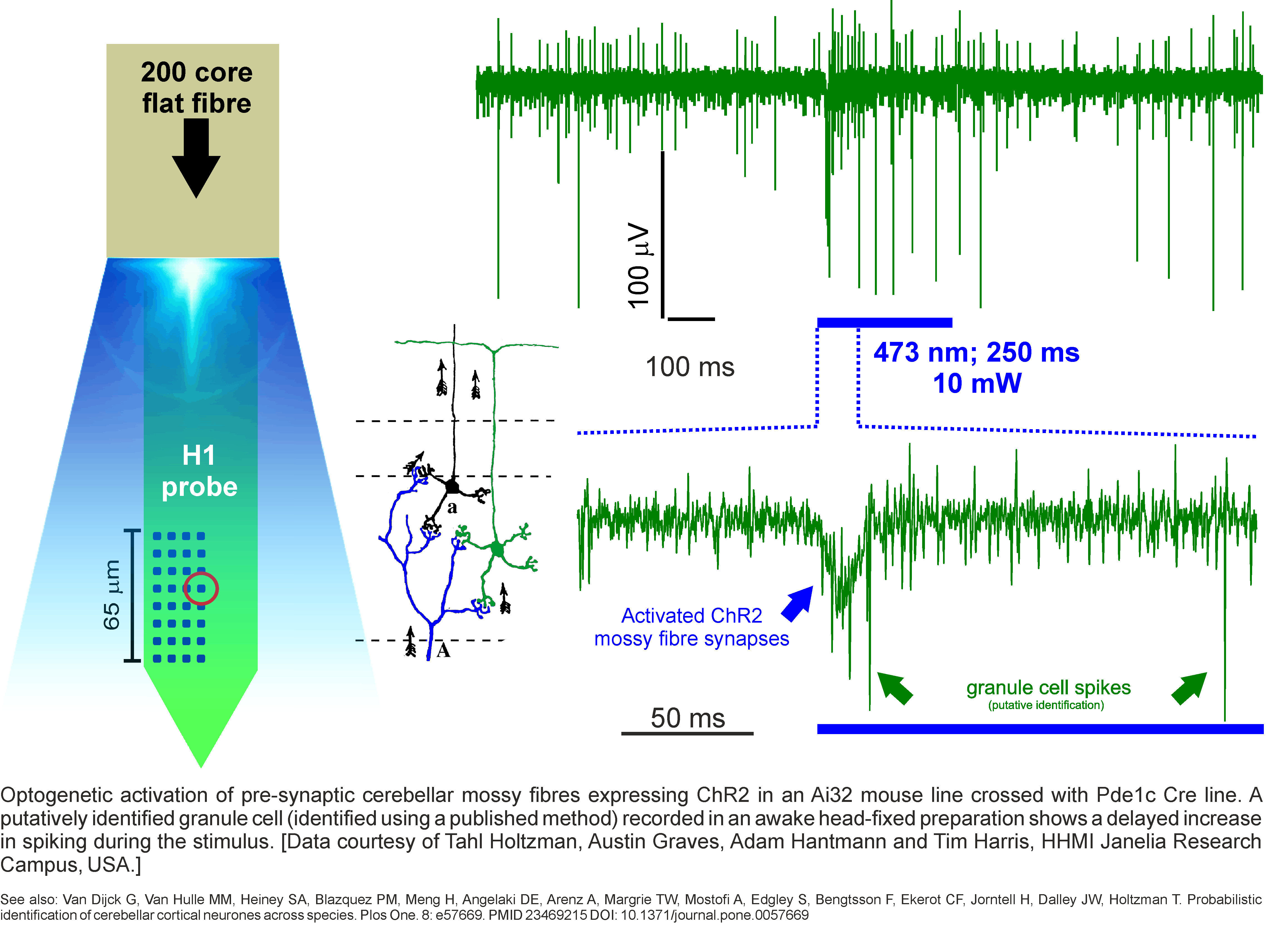
Combining in vivo optogenetics with electrophysiology is an important and versatile integration with broad relevance since it opens up the possibility to monitor single unit and local network activity during manipulation. This combined approach can be used for photo-identification of single units being recorded, interrogation of the contribution of defined inputs from one region to another to the circuit-level encoding of a stimulus or behavior and moreover for the establishment of closed-loop optogenetic manipulation based on fast-readout of network state / activity.

RELATED LINKS

Data examples using our acute and chronic optrodes:

Acute Optrodes - ready-made, reusable and minimally-invasive

Our ready-made acute optrodes are designed to be minimally-invasive with a defined (user-specified) spatial separation between our silicon neural probes and the fiber cannula to minimize local tissue damage, whilst maintaining proximity to spill sufficient light power to drive your chosen opsins. We offer a range of fibers to cater for brain surface illumination (200-core fiber) or deep-brain stimulation (100- or 60-core fiber) and our smaller diameter fibers also have angled 45-degree tips to aid with smooth movement through the tissue by minimizing tissue-drag and brain dimpling / compression.

Available with the following conventional fiber choices (1.25 mm ferrules):

  • 60-core; 0.37 NA; 45 degree angled tip | 100-core; 0.37 NA; 45 degree angled tip | 200-core; 0.66 NA; flat tip profile (recommended for surface-only illumination)

Taper-tip Lambda-B fibers for full-depth and dynamic-depth optrodes

Taper-tip Lambda-B fibers enable stimulation of large-volumes of brain tissue with a smaller, less-invasive footprint compared to conventional flat-tip fibers and without the need to move the fibers. It's also possible to dynamically stimulate at different depths and / or combining multiple wavelengths! Our innovative H-series and M-series silicon neural probes enable simultaneous single unit recording across multiple cell layers (e.g. cortex) so in combination with a taper-tip Lambda-B fiber you can now optogenetically stimulate and record across multiple cell layers. Moreover, since our probes are the only probes around designed to minimize photoelectric stimulation artifacts, you'll also enjoy clean, high-quality data.

Available with the following taper-tip Lambda-B fiber choices in collaboration with Optogenix (1.25 mm ferrules):

  • 100-core; 0.22 NA; 0.5/ 0.7 / 0.9 / 1.0 / 1.2 / 1.5 mm taper-tip | 200-core; 0.39 NA; 1 / 1.5 / 2 / 2.5 mm taper-tip | 200-core; 0.66 NA; 1 / 1.5 / 2 / 2.5 mm taper-tip

Ready-made acute optrodes - takes a closer look...

Acute-Neural-Probe-Optrode.gif#asset:670

Taper-tip-Lambda-B-fibers-for-whole-depth-and-dynamic-depth-optogenetic-stimulation.jpg#asset:754

Chronic Optrodes - Spatially-defined co-alignment of implantable fiber optics with silicon neural probes

Our chronic nano-Drives are designed to precisely co-align fiber cannulas alongside our silicon neural probes with spatially defined separation (300 or 650 microns between the fiber and probe) so as to minimize local tissue damage arising from the fiber, whilst maintaining proximity to the neurons being recorded to spill sufficient light power to drive your expressed target opsin. Our solution to the problem of uniting silicon neural probe microelectrodes with fiber optics allows both devices to be moved through the brain together in a freely-behaving animal, such that your electrodes are always in the right place with respect to your light-emission.

The chronic nano-Drives can also accommodate a fluidic injection guide-cannula alongside your optrode assembly - this facilitates an all-in-one approach whereby your chosen vector can be micro-injected at the end of your implant surgery via the guide-cannula, thereby guaranteeing that your optrode assembly will pass through your opsi-injected target. This innovative approach reduces mis-targeting errors within individual animals and makes redundant the traditional and error-prone approach of two surgeries; i.e. surgery (1) = injection and incubation time, surgery (2) = optrode implant by approximating the injection target location!


RELATED LINKS

chronic-optrodes.jpg#asset:1430

Chronic Probe Implant Protocol

Learn how to implant our chronic nano-Drives and probes (+/- fiber optics) to form compact, low-profile implants

Read More

Optrode assembly in action

A four-shank 64 channel probe with co-aligned fiber cannula smoothly penetrating the cortex [Courtesy of Maxym Myroshnychenko, Lab of Christopher Lapish, Indiana University - Purdue University Indianapolis, USA.]

Download our Catalog

Browse our complete range of tools; find your ideal probes and accessories

Read More